Sunday, June 29, 2014

Making an Ultrasound Trough

In the course of doing ultrasound training all over the country, I have been surprised to learn that equipment companies have been slow to accommodate the need for ultrasound troughs and tables for veterinarians.  Patient comfort during an exam makes everything go more smoothly. And, yes, you can always perform an exam with the patient in lateral recumbency, but you are still going to re-position multiple if you are thorough.

Many veterinarians use x-ray positioning troughs for ultrasound:  x-ray troughs were designed to hold a patient in position for a short time; they are too deep, too short, and too uncomfortable to maintain a patient in position for 15-20 minutes or longer.

We have made our own troughs from foam wedges covered with nylon pack cloth.  We use foam wedges that are standard cut and alter them to fit exam tables.  Our most commonly used wedge is 36” long, 16” wide, and 8” tall.  This size fits most exam tables in most clinics and since my practice is mobile, this is the trough we carry with us.  If your exam table can accommodate a longer trough, make yours longer.   If you are interested in making your own, tap one of your creative staff members or a family member and show that person the following:


To make an abdominal wedge V-trough support cushion for abdominal ultrasound patients you’ll need:  
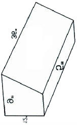
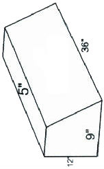
1. At least 2 foam wedges  5 x 9 x 12 x 36”  (3 if you would like to make your trough longer) order from:   http://www.foamforyou.com/back_support_wedges.htm#Back Bolsters if you don’t have a good source near you
2. Foam glue (spray can)wedge foam.jpg
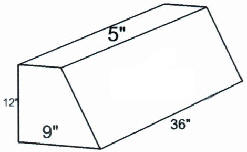
3. Cutting tool:  I like an electric knife if you can find one
4. Sharpie and yardstick

The wedge is going to be 16-18” wide depending on your exam table.  The 12” side of the wedge is the base. Using your yardstick and sharpie, draw a line 3.5 or 4 inchs from the 5” face to the angled face on both ends of both wedges.  Then connect the points on the the flat face and angled face.  Using the electric knife cut along the lines.  Now you have 4 wedges:  2 8 X 9 X 36” wedges and 2 4 X 7 (about) X 36” (keep the smaller pieces to make 1 or 2 smaller troughs for small dogs and cats)

Using the spray foam glue, follow the manufacturer’s instructions and glue the narrower faces of the two large wedges together to form a trough.  Glue the two smaller pieces together, as well.

5.    Now you have two troughs.  Use large garbage bags to wrap your troughs before covering for waterproofing (I know people who stop here and simply cover the garbage bag wrapped foam with towels!)
wedge foam 1.jpgwedge foam 2.jpg
IF you want to make a cover:

1.  Nylon pack cloth or other densely woven, water resistant fabric
2.  Heavy duty thread
3.  1.5 yards sew on 2” velcro (don’t use self stick velcro)
4.  Sewing machine
5.  Butcher paper to make a pattern
6.  Someone who understands how to make a pattern, cut and sew!

Friday, April 20, 2012

Pulmonary Hypertension

Cardiac ultrasound can be used to show the presence of pulmonary hypertension if there is a tricuspid insufficiency, pulmonary stenosis, or pulmonary insufficiency.  Continuous Wave Doppler is used to determine the velocity and pressure gradient of the valvular flow.  Pulsed Wave Doppler is not capable of measuring higher flow rates.  Pressure gradient indicates the difference between the pumping chamber (the right ventricle in this case) and the receiving chamber (either the right atrium in tricuspid insufficiency or the pulmonary artery in pulmonary stenosis).  A pressure gradient of 10-50 mm Hg is considered to mild pulmonary hypertension, 50-80 mm Hg is moderate, and greater than 80 mm Hg is severe.

Radiographs can also be used to show more severe pulmonary hypertension.  If the anterior pulmonary arteries are larger than the veins on the lateral views, pulmonary hypertension is present.  On the VD or DV views, the caudal pulmonary arteries will be larger than the veins.     

There seems to be some confusion about treatment of pulmonary hypertension.  Some recent lectures have unintentionally given the idea that all cases must be treated and that Viagra is the treatment.  First, show that pulmonary hypertension is present.  Then, identify and treat the cause of the hypertension.

If heartworm disease is present, treat the heartworm disease.  If pneumonia is present, treat the pneumonia, etc.

If pulmonary valve stenosis is present, mild cases (PG of 10-50 mm Hg) are not treated.  The patient will probably live a full life without symptoms or need for treatment.  In moderate to severe cases, ballon valvuloplasty can be done to partially relieve the stenosis.

Pulmonary hypertension is a secondary problem with mitral insufficiency or mitral stenosis because blood backs up to the lungs.  The right ventricle must pump against the increased resistance of the mitral insufficiency.  The presence of a tricuspid insufficiency allows us to use Continuous Wave Doppler to estimate the pressure of the right ventricle.  Normal pressure in the right ventricle is 20 mm Hg and a pressure of 30 mm Hg or higher is considered to represent pulmonary hypertension.  Usually, treatment of the mitral insufficiency will also treat the pulmonary hypertension.  

Viagra is usually only used in the last 30 days of the patient's life when end stage disease results in uncontrollable pleural effusion.  This can be seen in , end stage heart disease (severe mitral insufficiency, tricuspid insufficiency, pulmonary stenosis, pulmonary insufficiency, aortic stenosis, aortic insufficiency), pulmonary fibrosis, chronic fungal pneumonia, neoplasia, or other infiltrative diseases.

Saturday, February 18, 2012

What Kind of Machine Should I Buy 3

Next, look for a machine that is user friendly. How can you tell if it will be easier to use? Look at the keyboard. Are the buttons, knobs, or toggles clearly labeled as to the function they control? With some machines, each button, knob, or toggle may have multiple functions. Ask to see the owner’s manual. If there is one booklet, the machine is probably user friendly. If there are 3 or 5 separate booklets, it is a very complicated machine. In my opinion, the complicated, “big box” ultrasound machine is capable of making multiple adjustments to give the absolute best quality image, but the operator must know and use all of the available button, knobs, and toggles to get that image. This type of machine may cost $80,000 to $350,000 or more and is a necessity for a radiologist or cardiologist. It is perfectly acceptable if this is the kind of machine you want. In my opinion, this is more ultrasound machine than a general practitioner needs. In my opinion, a general practitioner needs a machine that is user friendly and can be quickly learned and put to use in the practice. In my experience, when machines or procedures are difficult or tedious, most people will not use the machine or do the procedures. Take the time (2-3 days) to study the owner’s manual and learn how to use the machine you buy.

Fourth, what kind of training is available to you? An ultrasound machine is worthless if you do not know how to use it. Some ultrasound sales companies include training in the cost of the machine. It is not free. You are paying for it whether you use it or not. You will probably have to pay for your own travel and lodging to get to the training. You may consider separating the cost of training from the cast of the machine, especially if you are financing the cost of the machine. Some ultrasound companies offer training for a fee.

Saturday, February 11, 2012

What Ultrasound Machine Should I Buy Part 2


After you have determined what your practice needs are, as you research ultrasound machines available, ask if the machine is capable of supporting the procedures you want and how many transducers you will need to do those procedures.  If you start with abdomens but eventually want to do hearts as well, does the machine have cardiac capability or can it be upgraded to cardiac functions when you are ready?  Sometimes you just need to buy a cardiac transducer when you are ready.  Sometimes you have to buy a new machine that can do hearts and abdomens.  Think about your future needs.  For shallow depths (like cat or exotic abdomens, equine tendons or repro) you need higher frequency transducers.  For deeper depths (like large dog or equine abdomens) you need lower frequency transducers.  For most procedures in the small animal abdomen, you want a micro-convex transducer.  For shallow structures (pancreas, intestines) a linear transducer could be helpful.  For cat hearts you need a higher frequency cardiac transducer.  For some large dogs and horses, you need a lower frequency cardiac transducer.  Colorflow Doppler is very useful in the abdomen, but it will increase the cost of the machine.  For cardiac studies, Colorflow and Spectral Doppler (both Pulsed Wave and Continuous Wave) are necessities and will increase your total cost.  I caution you not to buy a machine because it is cheap, but because it will do what you need.    For diagnostic purposes in any heart, you MUST HAVE COLORFLOW DOPPLER, PULSED WAVE DOPPLER, AND CONTINUOUS WAVE DOPPLER.  Will the machine allow you to easily download patient information and images?  Can it do video clips?  Can the hardware and software be upgraded?  How long is the warranty?  If it is an older model, how much longer will the manufacturer support repairs?    

Tuesday, February 7, 2012

Phantom

Someone just asked how to make a practice phantom since commercially available phantoms are rather expensive and have limited usage life.  When training, I use a jello mold filled with objects (mostly dried fruit) for developing scanning and fine needle biopsy skills.  It is a homemade phantom that is inexpensive and effective.

My Recipe: 

2 Large Packages Black Cherry Jello (the darkest color available to disguise the objects you add) + 2 packets of plain Knox gelatin (for firmness)
Disolve Jello and gelatin in 3 cups of hot water (not boiling)
Pour mixture into a 1 quart ziploc freezer bag.  Add 12 prunes.  Eliminate as much air as possible as you seal the bag.  Lay the bag flat in the refrigerator and allow the gelatin to set.  It will become quite firm.  Remove from the refrigerator, use ultrasound gel, and practice.

Sometimes I add small balloons filled with milk or water for cystocentesis practice.

Many people figured out how to use Jello to make phantoms.  I adapted my recipe from Knox Blox.  How many of you are old enough to remember those?  I think, more recently, they're called Jigglers.

Saturday, February 4, 2012

What Kind of Machine Do I Buy?


What kind of ultrasound machine should I buy?
Many people visit our booth at veterinary convention exhibit halls to ask this question.  It is a very good question but will take some time to give a good answer.  I will divide this into four topics:

1.       List the ultrasound procedures you want to do
2.       Search for a machine that will support the applications you want to do
3.       Look for a machine that is user friendly
4.       Ask what kind of training is available 

Assess your wants and needs:   you need to decide what kind of ultrasound procedures you want to be able to perform BEFORE you start researching equipment.  If your practice is limited to small animals, do you want to limit yourself to abdominal ultrasound only, or would you like to expand to cardiac ultrasound some day?  Are you interested in exotic animals or reproduction studies?  If you are limited to large animal practice, do you want to do tendons, reproduction, cardiac, and abdominal studies?  If you are in a mixed animal practice, do you want to do everything?  Make a list of the types of procedures you want to be able to perform.  If you have multiple doctors in your practice, enlist them in the decision making process. 

 WHY?  The cost of your unit will depend upon the requirements you place on it. Your basic unit is hardware consisting of a computer processor (also known as the platform) attached to transducers (which gather the information that feeds the processor and a screen (which displays the images generated by the processor).  Most new ultrasound units display very good to excellent images.  Transducers, though versatile, tend to be task specific so it is important to know what tasks you want to perform.   The platforms depend upon software written to accomplish specific tasks:  creating an image, measurement and labeling packages, filing, retrieving data etc.  If your facility is computerized, you may also want to ask if the units you are considering are easily compatible with your current image storage system.
So, when looking for an ultrasound unit, it is very important for you to have a list of the types of procedures you want to perform, the types of patients you serve in your practice, and the kind of record storage you use in your clinic or hospital.